The shoulder joint has a rich arterial supply via several branches of the subclavian and axillary arteries to safeguard against ischemia.
This model started as a rough segmentation of the bones in the right shoulder. The data was segmented from a CT scan in the D2P software. From this segmentation, the bones were smoothed, and a large fracture in the head of the humerus was repaired in Pixologic Zbrush. The ligaments of the shoulder, and the arterial supply were modeled de novo in Zbrush. Materials and final lighting and rendering were completed in Zbrush.
Animation
Shining a Light on Phototropism
This animation started as an original script and storyboard. The modeling, texturing, lighting, and rendering were completed in Cinema4D. Post effects were applied in Adobe After Effects.
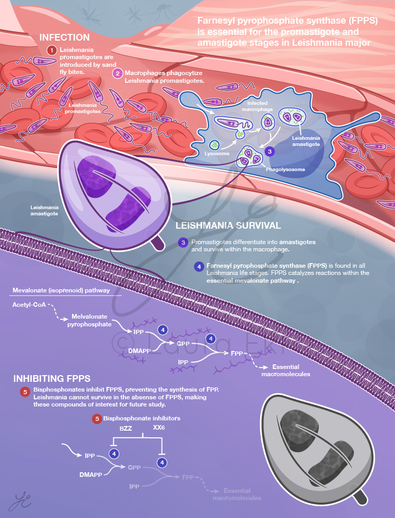
This enzyme catalyzes two critical steps in the mevalonate pathway, producing the precursors to essential molecules in eukaryotes. This enzyme is critical to survival of both the amastigote and promastigote stages in Leishmania.
The molecular structure of this model was retrieved from PDB and visualized in Cinema4D via the ePMV plugin.
Have a project in mind?
I’m currently accepting freelance illustration work.